Anatomy Muscles Pelvis / Your Pelvic Floor Muscles Yamuna. It is composed of three separate paired muscles; (2) the levator ani and the coccygeus, which together form the pelvic diaphragm and are associated with the pelvic viscera. This mri male pelvis axial cross sectional anatomy tool is absolutely free to use. We'll look at these structures first in a male specimen. •sacrumandcoccyx, which form the posterior wall.
(1) the obturator internus and the piriformis, which are muscles of the lower extremity, and will be described with these (pages 476 and 477); Muscles that attach from the pelvis to the trunk and cross the lumbosacral joint muscles that attach from the pelvis to the thigh/leg and cross the hip joint pelvic floor muscles that are located wholly within the pelvis Sacrotuberous ligament on either side.; •sacrumandcoccyx, which form the posterior wall. The structure of the pelvis supports the contents of the abdomen while also helping to transfer the weight from the spine to the lower limbs.
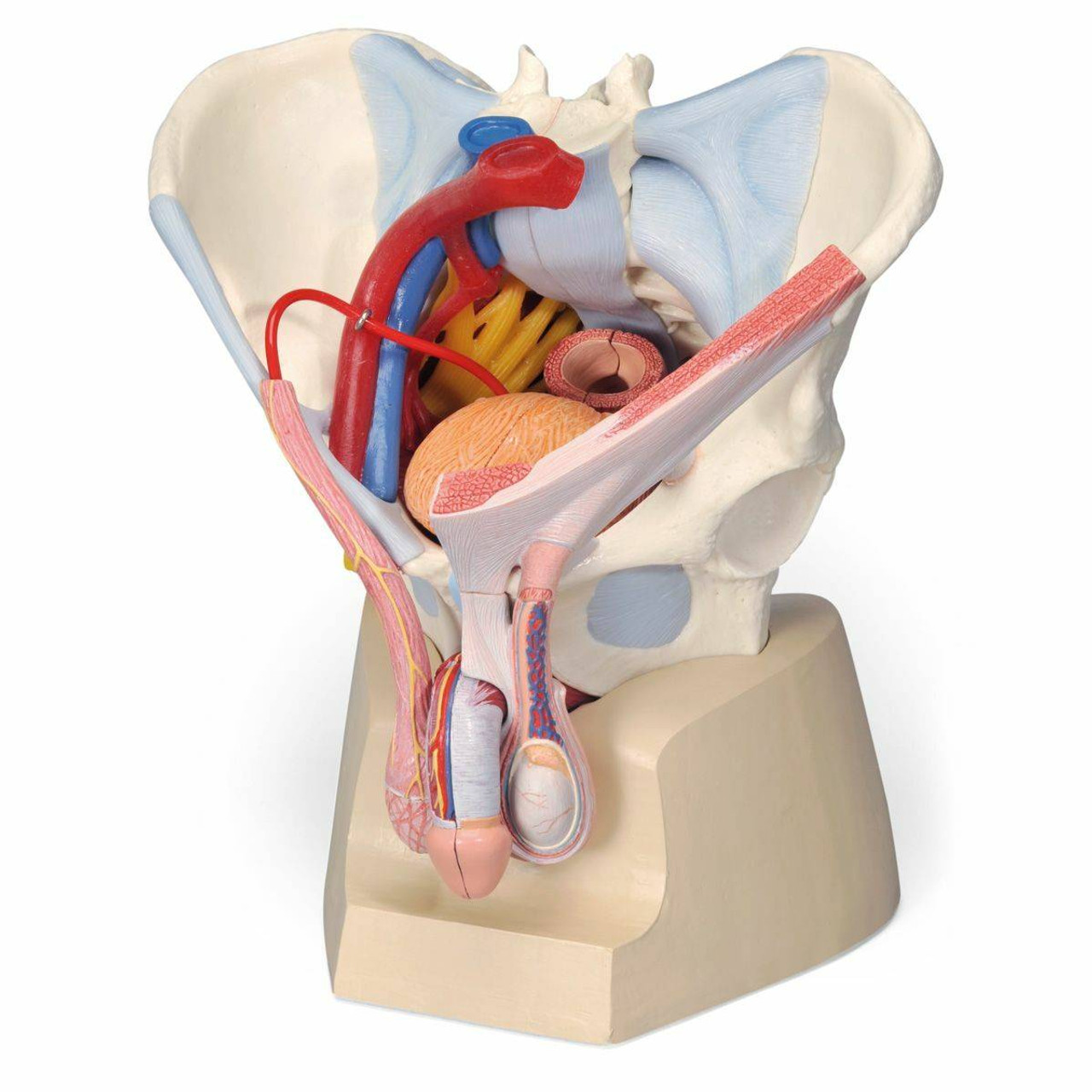
Anatomy Model Male Pelvis Ligaments Organs from cdn11.bigcommerce.com (2) the levator ani and the coccygeus, which together form the pelvic diaphragm and are associated with the pelvic viscera. The muscles of true pelvis are: It's supplied by ventral rami of first and 2nd sacral nerves (s1, s2). The iliopsoas muscle consists of the iliac muscle, which comes from the inner surface of the ilium in the pelvis, and the psoas muscle, which originates from the vertebral column. ▪the bony pelvis is composed of four bones: (1) the obturator internus and the piriformis, which are muscles of the lower extremity, and will be described with these (pages 476 and 477); The muscles of the pelvis and hip control the vast range of movement of the legs and torso. These muscles have attachments to the pelvis as follows:
A proper kegel exercise means a full contraction and relaxation of the pc muscle.
Muscles an important group of muscles in the pelvis is the pelvic floor. ▪the bony pelvis is composed of four bones: They form a large sheet of skeletal muscle that is thicker in some areas than in others. These muscles originate near the anteroinferior external surface of the bony pelvis and insert at the linea aspera. This mri male pelvis axial cross sectional anatomy tool is absolutely free to use. The floor of the pelvis is formed by the two muscles named levator ani and coccygeus. The levator ani muscles are the largest group of muscles in the pelvis. The iliopsoas muscle consists of the iliac muscle, which comes from the inner surface of the ilium in the pelvis, and the psoas muscle, which originates from the vertebral column. The four groups are the anterior group, the posterior group, adductor group. Rectus femoris muscle, one of the quadriceps muscles on the front of your thigh. (2) the levator ani and the coccygeus, which together form the pelvic diaphragm and are associated with the pelvic viscera. These muscles have attachments to the pelvis as follows: Piriformis the piriformis is a triangular muscle 1 on either side on the very front of the posterior wall of true pelvis.
They form a large sheet of skeletal muscle that is thicker in some areas than in others. Piriformis the piriformis is a triangular muscle 1 on either side on the very front of the posterior wall of true pelvis. Study human anatomy with reliable 3d models & detailed articles. The pubococcygeus (pc) muscle is the muscle that runs the show in pelvic floor health. The many muscles of the hip provide movement, strength, and stability to the hip joint and the bones of the hip and thigh.
The Pelvic Floor Pelvic Floor Yoga Anatomy Kegel Exercise from i.pinimg.com The muscles of true pelvis are: Arcus tendineus levator ani and the ischial spine Piriformis the piriformis is a triangular muscle 1 on either side on the very front of the posterior wall of true pelvis. We'll look at these structures first in a male specimen. The muscles of the pelvis and hip control the vast range of movement of the legs and torso. The levator ani muscles consist of three. These muscles originate near the anteroinferior external surface of the bony pelvis and insert at the linea aspera. It can be described as one of the bodies diaphragms.
It's supplied by ventral rami of first and 2nd sacral nerves (s1, s2).
Then we'll look at the complex sheet of muscles, collectively called the pelvic diaphragm, which form the floor of the pelvic cavity. The hip joint is one of the most flexible joints in the entire human body. The pelvic inlet involves three of the four units of which the bone pelvis is composed. The pubococcygeus (pc) muscle is the muscle that runs the show in pelvic floor health. Psoas consists of a pair of deep muscles (psoas major and iliacus) located on each side of the pelvis in the abdomen. The iliopsoas muscle consists of the iliac muscle, which comes from the inner surface of the ilium in the pelvis, and the psoas muscle, which originates from the vertebral column. The muscles of the pelvic floor are collectively referred to as the levator ani and coccygeus muscles. These two muscles join each other and then attach to the lesser trochanter. The medial surface provides attachment for both transverse perinei, obturator internus and externus, piriformis, coccygeus and levator ani muscles. It's supplied by ventral rami of first and 2nd sacral nerves (s1, s2). It can be described as one of the bodies diaphragms. These muscles have attachments to the pelvis as follows: The muscles within the pelvis may be divided into two groups:
These muscles can be grouped based upon their location and function. Pelvis anatomy muscle thigh muscular system pelvis text hand human png pngwing from w7.pngwing.com this mri pelvis cross sectional anatomy tool is absolutely free to use. •sacrumandcoccyx, which form the posterior wall. The pubococcygeus (pc) muscle is the muscle that runs the show in pelvic floor health. Muscles that attach from the pelvis to the trunk and cross the lumbosacral joint muscles that attach from the pelvis to the thigh/leg and cross the hip joint pelvic floor muscles that are located wholly within the pelvis
Blood Vessel Anatomia Y Fisiologia Muscle Pelvis Nerve Pelvic Orange Anatomy Human Body Png Klipartz from c0.klipartz.com Ligaments, tendons, and muscles play an important role in the function of the hip. The pelvis consists of the sacrum, the coccyx, the ischium, the ilium, and the pubis. Muscles play an important role in the. The thigh bone or femur and the pelvis join to form the hip joint. The four groups are the anterior group, the posterior group, adductor group. Rectus femoris muscle, one of the quadriceps muscles on the front of your thigh. These muscles arise from the hip, spine, and proximal femur. The pubococcygeus (pc) muscle is the muscle that runs the show in pelvic floor health.
These two muscles join each other and then attach to the lesser trochanter.
The muscles of true pelvis are: The muscles within the pelvis may be divided into two groups: Pelvis anatomy muscle thigh muscular system pelvis text hand human png pngwing from w7.pngwing.com this mri pelvis cross sectional anatomy tool is absolutely free to use. The levator ani is a broad sheet of muscle. These muscles originate near the anteroinferior external surface of the bony pelvis and insert at the linea aspera. The pelvic floor muscles provide foundational support for the intestines and bladder. These two muscles join each other and then attach to the lesser trochanter. It can be described as one of the bodies diaphragms. These muscles all serve as adductors of the thigh, but also serve as important stabilizers of the pelvis and work to maintain balance of the pelvis on the lower limb during gait. A proper kegel exercise means a full contraction and relaxation of the pc muscle. Muscles an important group of muscles in the pelvis is the pelvic floor. Explore every muscle, bone and organ in 3d They are responsible for bending and adducting the thigh.